Getting closer to co-localised proteins!
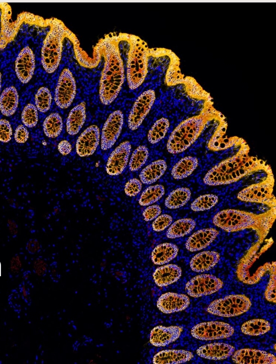
Multiple labelling on tissues and cells using immunohistochemistry or immunofluorescence can have challenges in any way, but especially when trying to demonstrate co-localised proteins.
Colorimetric substrates are usually only used where antigens are at least in different cell compartments because the deposition of the substrate can hinder the access of subsequent reagents to their target.
For immunofluorescence, the staining can be closer; however, the resolution of regular microscopes might not be sufficient to demonstrate conclusively if proteins are close enough to interact.
Proximity ligation assays (PLA) are a great way to demonstrate antigens located within 40 µm of each other—at the very limit or beyond the capability of current supermicroscopy techniques—with just a regular-resolution microscope!
The principle of the assay is that both of your antibodies need to be close enough together to generate a signal with a rolling circle amplification, detected with either fluorescence or colorimetry. Kits are available to detect combinations of mouse, rabbit, and goat antibodies on tissues or cells, as well as ready-to-use kits for essential target combinations like PD1/PD-L1.
If you’re multiple labelling already, it’s likely you’re already most of the way there with your optimisation for PLA!
See the range of flexible kits here, or request a consultation with our in-house IHC expert if you would like to find out more!