Blocking Reagents
Vector Laboratories has a range of blocking reagents to prevent background interference from different sources.
Jump to...
Vector®TrueVIEW™ Autofluorescence Quenching Kit
Reveal true immunofluorescence—even in the most challenging tissues
In tissue sections, autofluorescence is the unwanted fluorescence that can make it difficult or impossible to distinguish antigen-specific signal from non-specific background. The novel, patent-pending Vector®TrueVIEW™ Autofluorescence Quenching Kit specifically binds and quenches autofluorescent elements from non-lipofuscin sources, significantly enhancing signal-to-noise in most immunofluorescence assays.
Why TrueVIEW™ Quencher?
- Specific reduction of autofluorescence from non-lipofuscin sources
- Easy-to-use, one-step method
- Quick 5 min incubation
- Compatible with a wide selection of fluorophores
- Compatible with standard epifluorescence and confocal laser microscopes
- Fluorescence assays
- Vector TrueView kit contains VECTASHIELD Vibrance Mounting media for fluorescent signal preservation
- Quenches autofluorescence caused by aldehyde fixation, and on problem tissues such as liver, spleen, red blood cells & kidney.
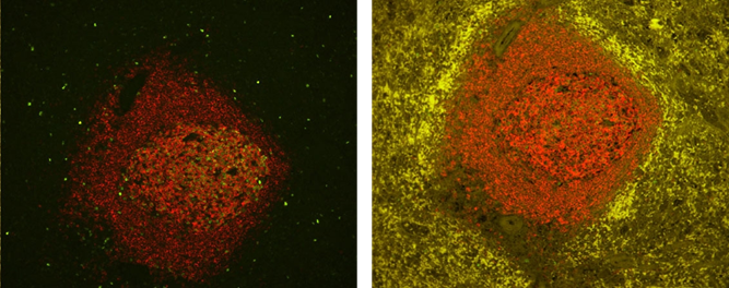
 Without TrueView vs With TrueView
Reduction of autofluorescence in human spleen using the TrueVIEW™ Autofluorescence Quenching Kit. Human spleen sections (FFPE) stained using mouse anti-CD20 (red) and rabbit anti-Ki67 (green) primary antibodies detected with VectaFluor™ Duet kit (DK-8818). Note significant reduction of autofluorescence in the treated section.
Comparison Between Vector® TrueVIEW™ Reagent and Other Autofluorescence Reducing Agents
We compared the effectiveness of Vector TrueVIEW quenching action in parallel with other commercially available autofluorescence reducing products and "home brew" reagents, on serial sections of formalin-fixed, paraffin-embedded human pancreas visualized using a standard fluorescein (green) filter. No specific immunofluorescence staining was conducted. The images below highlight our results. All images were acquired under identical conditions (including microscope objective and exposure times).
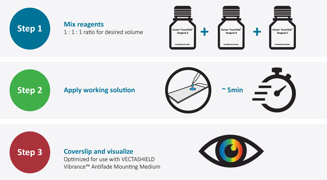
Easy to Use
Following completion of immunofluorescence staining:

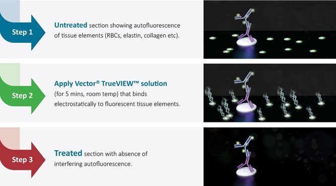
Mode of Action
Following completion of the IF staining procedure:

Protein Blocking Solutions
Non-specific protein interactions between detection reagents and target protein elements can cause unwanted background staining. To minimize this type of interaction, use an appropriate protein blocking reagent:
- Normal sera — widely used as a general protein block in tissue and cell-based staining. For best results, choose a serum species for blocking that matches the species of the secondary antibody.
- Carbo-Free Blocking Solution (SP-5040) — for lectin staining or in multiplex IHC applications. Lectins are used for at least one label because this solution does not contain interfering glycoproteins.
- BSA (SP-5050) — free of the glycoproteins and impurities present in other grades of BSA that can introduce artifacts or increase background staining. Use in IHC, iCC, IF, blots, and ELISAs.
- R.T.U. Animal-Free Block and Diluent (SP-5035) — plant-protein derived universal antibody diluent and protein blocking reagent for use in multiplex IHC applications where primaries or secondaries from multiple species are used. This solution does not contain animal immunoglobulins, which may be non-specifically bound by the secondary antibodies.
Prostate without and with R.T.U. Animal Free Block and Diluent used in the blocking step. Both sections are no primary antibody controls with Biotinylated Universal Secondary Antibody, VECTASTAIN® Elite® ABC, and ® Vector DAB Substrate for detection. Note significant toning in section without RTU Animal Free Block and Diluent.
Western Blot Blocking Solutions
In dot blot or western blot application, blocking solutions minimize the non-specific binding of detection antibodies to the membrane that lead to background.
Vector Laboratories offers two solutions formulated to block membranes in blotting applications:
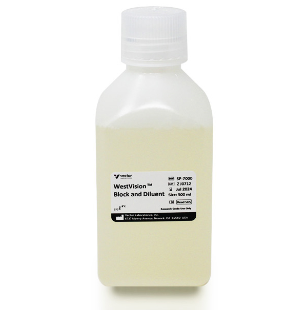
- WestVision™ Block and Diluent — a proprietary, ready-to-use formulation containing Tween 20 that improves signal intensity and resolution without increasing background. It is compatible with both alkaline phosphatase- and peroxidase- based detections systems, and with both chemiluminescence and chromogenic development.
- Animal-Free Blocker™ — a plant-derived universal antibody diluent and blocking reagent for nucleic acid and protein blotting, IHC, and for other applications. It contains no animal-derived protein and can be used as an alternative to sera, BSA, casein, or non-fat dry milk.
4-fold serial dilutions of human recovered plasma were transferred to nitrocellulose. Membranes were blocked with WestVistionTM Block & Diluent (A) or Competitor Block (B), probed with Rabbit anti-Transferrin antibody (0.25 ug/ml), and detected with WestV.
